Imaging in Neuroscience
Already 500 years ago researchers were fascinated by the workings of the brain. Leonardo da Vinci, who used phrenology to investigate the black box of the brain, was still denied deeper insights. Only since the rise of modern medical imaging techniques has it been possible for researchers to take a "look" into the brain of a living person. These modern methods usually use radiation, magnetic fields or ultrasound to visualize the functioning of the brain. Imaging techniques can be used in many fields of application. For example, neuroimaging techniques are also used in the legal system when it comes to assessing the culpability, recidivism prognosis or credibility of criminals. This status report focuses on the application and ethical aspects of its use in medicine and, in particular, in medical practice.
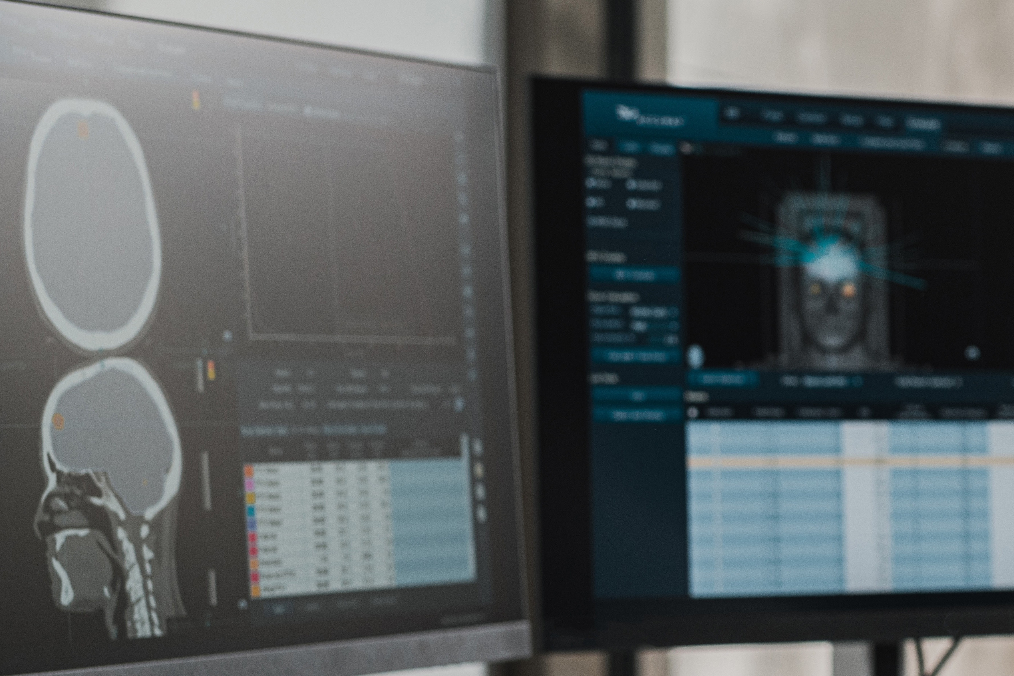
What are imaging techniques?
Imaging techniques are a neuroscientific tool used to contribute to improved diagnostics and therapy planning, for example prior to surgical interventions. Imaging techniques now also help researchers to answer fundamental questions about the structure and composition of the brain - a new possibility that is in particular due to magnetic resonance imaging (MRI), as it does not involve harmful radiation and can therefore be reproduced multiple times without any concerns.
Computed tomography, or CT for short, can provide information particularly in clinical diagnostics and acute changes in the brain, such as blood clots or strokes, and is still considered the gold standard despite the use of X-rays.
Magnetoencephalography (MEG) and electroencephalography (EEG) are also non-invasive measures. In MEG procedures, around 300 sensors are distributed over the entire surface of the head, enabling neuronal activity to be recorded in the millisecond range. This ensures an undistorted picture of brain activity, particularly in the case of MEG. Similar to EEG, near-infrared spectroscopy (NIRS) also derives signals directly from the surface of the skull. The advantage of NIRS is that it is easy to handle and can also be used on the move. This also provides an imaging method that can deliver results in everyday situations away from sterile laboratory conditions. Although NIRS offers a lower resolution, mobility is a decisive argument for its use in some situations.
However, it is important to understand that the term "imaging" does not actually mean the depiction of a clear picture of the internal processes of the brain, but rather uses data to evaluate which areas are active or whether, for example, tumors or hemorrhages are present in the brain.
Controversy surrounding the recognition of neuroimaging in the sciences
While neuroimaging has made a significant contribution to our understanding of the brain over the last 30 years, this field of research also initially had to deal with inadequate results and the problem of insufficient sample sizes and a lack of reproducibility. However, while the number of test subjects was initially around 10 per group, this size quadrupled by the 2000s and has been significantly increased again in the last 10 years. Thanks to the paradigm shift towards global data sharing and the exchange of collected data worldwide, researchers now have access to a large pool of imaging data ranging from MRI to EEG.
This data and the information gained from it has enabled further advances in the study of brain connectivity and ageing processes, as well as the relationship between brain structure and environmental factors. With the help of large databases such as the UK Biobank Cohort or the 100 Brains Study, for example, the default mode network could be determined and a connection between this network and the natural ageing process could be identified. These databases can also be used to determine how protective and negative lifestyle factors affect the brain structure and functional connectivity of the brain. Here it was possible to recognise that negative factors such as excessive alcohol consumption and excessive smoking, but also excessive sport, lead to a regional loss or loss of connectivity of the synaptic connections, while social integration in particular contributes to the preservation of these.
Possible applications of neuroimaging
Now that imaging procedures are widely recognised as a helpful measuring instrument, these procedures, especially neuroimaging procedures such as CT and MRI, are primarily used in psychiatric and neurological diagnostics. The data obtained from these procedures can provide prognostic support, diagnose acute psychiatric and vascular diseases and thus be decisive for treatment. Now that the human genome has been decoded, work is underway to break down the human brain and decode its functions - but science is still a long way from achieving this. Nevertheless, it is already possible to identify the first markers for Alzheimer's dementia based on the deposition of amyloid plaque or to detect bleeding and tumors in the brain. Research into psychiatric illnesses such as psychosis is also on the way to identifying markers for better diagnosis and early detection. Nevertheless, it is important to remember that the human brain does not exist independently but is an organ that is constantly interacting and changing with its environment. The greatest strength of the brain is certainly its inherent learning ability and flexibility, which enables people to prevent degeneration processes and to establish new neuronal connections even later in life.
The potential of neuroimaging in medical practice is illustrated in more detail below using two examples.
Example of the diagnosis of acute stroke
Imaging techniques have long been used to identify strokes, whereas CT and MRI scans can also be used to define bleeding, vascular occlusions and the size of an infarct core. The identification of the vascular occlusion responsible for the stroke is crucial for further clinical treatment. This is particularly determined by using CTA and MRA. These technologies are both fast and efficient and provide an image of the arterial system of the entire body in the shortest possible time. It is precisely the time factor that often plays a significant role in infarcts - diffusion MRI has proven its worth here, providing a good prognosis in the early detection of infarct cores. MRI is also used in patients whose onset of infarction is undetermined, for example because they were asleep. The diffusion/perfusion mismatch is also decisive here. After determining this imbalance in the MRI sequences, clinical treatment can be adjusted and, for example, intravenous thrombolysis can be indicated to dissolve the blood clot.
Today, both CT and MRI provide important prognostic and diagnostic information that helps determine the therapeutic approach. Imaging is still irreplaceable in acute stroke cases.
Imaging procedures can also provide information on long-term recovery, particularly when it comes to the prognosis of recovery. Structural MRI is used here, along with other factors. These indicators, together with the localisation of the brain damage, allow reasonably valid prognoses to be made. Nevertheless, even these findings from CT and MRI are not sufficient to determine a final prognosis for recovery with certainty. The prognosis can only be improved by adding functional biomarkers. Overall, lesions in the brain can be identified using MRI procedures, DWI and DTI map the structural connectivity of certain areas of the brain. Resting-state fMRI depicts functional connectivity in network regions, while activation fMRI contrasts the regions that should be primarily involved in a corresponding function, regardless of lesions.
As described, MRI and CT are the most important imaging tools for recognizing a stroke and drawing both diagnostic and prognostic conclusions. MEG and EEG can also be consulted here but play a subordinate role at best.
Example of imaging procedures for neurodegenerative diseases
Imaging procedures are also consulted in neurodegenerative procedures when it comes to making a diagnosis. Until recently, these procedures were only used for differential diagnosis, but today we are getting closer to biomarkers and a valid interpretation. Nevertheless, it is still difficult, even for specialized professionals, to make a diagnosis of Parkinson's disease (PD), dementia with Lewy bodies (DLB), multisystem atrophy (MSA), progressive supranuclear palsy (PSP) or corticobasal syndrome and to separate them from other diseases. New measurement techniques such as high-field and ultra-high-field MRI, which enable faster image acquisition, as well as additional MR sequences that image neuromelanin and iron, are helpful in this regard. The combination of molecular imaging and the acquisition of MR sequences makes it possible to read out dysfunctions of dopaminergic (dopamine), serotonergic (serotonin), cholinergic (acetylcholine) and noradrenergic (noradrenaline) processes in the brain and thus provides information about a possible early diagnosis of Parkinson's disease. In the future, it is expected that higher field strengths in the measurement of MR sequences will allow even better, more valid results, and MEG recordings are also expected to be used more widely in the diagnosis and treatment of neurodegenerative diseases.
In the examples described above, it is essential to provide an ethical framework that medical and scientific professionals can fall back on when it comes to issues such as research studies, incidental findings or even the indication of an imaging procedure. The autonomy of patients and test subjects must always be a top priority and coercion of the person concerned must be avoided. Sociological factors such as background or milieu also play a role here, as studies can, for example, expose low-income test subjects to indirect coercion.
Incidental findings also pose an ethical problem for doctors: the indicated diagnostic methods are not congruent with the intended findings, but it is difficult to ask the patient for consent for an incidental finding afterwards, which presents healthcare professionals with the dilemma of preserving life versus the patient's autonomy. A more basic, albeit equally important, challenge is weighing up an imaging procedure against the pain and possible psychological stress that such a procedure entails. Here, the attending physician is instructed to carry out a risk-benefit analysis and weigh up whether the benefit in terms of a cure outweighs the risk of pain or deterioration of the patient's state of health.
In ethics, the imaging procedures described above are also summarized under the term neuroimaging. However, the meaning is the same. Neuroimaging or imaging procedures entail a number of risks and ethical implications, which are discussed below and which must be considered in order to ensure safe, regular use on test subjects and patients without reservation.
Risks of use
Imaging procedures offer many possibilities, but also entail a number of risks, some of which are minimal, but some of which are more serious due to radiation exposure. For example, NIRS, EEG and MEG carry hardly any risks, while the risk of MRI is considered to be slightly higher and that of PET and SPECT significantly higher. In the case of MRI procedures, one of the greater dangers is the magnetic field emitted by MRI scanners. Patients may unknowingly introduce magnetic objects into the MRI device - from magnetic sand to magnetic color pigments in tattoos, there are many objects that can be unintentionally introduced into the device. Older implants also pose a risk here, as they are also magnetic and therefore not suitable for MRI devices. In addition to the negligent insertion of magnetic objects, the high noise level and the confined space of the MRI machine are also a potential risk, which can be psychologically and physically stressful for people with claustrophobia. The administration of contrast agents, which in most cases consist of substances containing iodine or gadolinium, can also lead to such serious negative consequences in the rare case of side effects that their administration must always be assessed in terms of a risk-benefit balance.
Nevertheless, MRI is still considered a minimally risky procedure, unlike SPECT and PET. As radionuclides are used in both procedures, they carry a greater risk in themselves. Users are exposed to the radiation during handling, while patients are injected with the substances - so there is a twofold risk on the patient side: the invasive procedure can expose the body to alpha and beta radiation, and the injected agent means that the body is also exposed to an internal radiation source.
When it comes to the question of age specification in the application, it can be stated that the use of imaging procedures on children can only be justified to a limited extent - while some researchers consider the playful involvement of children in research with these procedures to be justified if a correspondingly large gain in knowledge is created for individuals or groups, other researchers reject this with the thought of direct suffering for the respective subjects.
The challenge of providing appropriate information to subjects and patients
Transparent and successful communication with subjects and patients must also be considered. For example, it should be explained that the imaging procedures are not images in the true sense of the word, but rather measured values that are color-coded. Secondly, there are often misunderstandings about what the individual results are being compared with: For example, the measured values of a patient are not compared with individual samples of another individual, but with the average of a group of test subjects - deviations are therefore the rule. Even with the best information, misjudgments often continue to occur, especially on the part of the patient. Expensive procedures often promise greater benefits for recovery and patients are disappointed when the procedure does not offer any new chances of treatment or recovery. This phenomenon is also known as diagnostic-therapeutic misjudgment: a therapeutic benefit is estimated to be significantly lower or, more often, significantly higher than it actually is.
Another widespread misunderstanding in the practice with imaging procedures is the therapeutic misunderstanding. This refers to the fact that participants in a study do not directly benefit from it and that no cure is intended. In such studies, an improvement in health is more of a side effect that may occur - but the study is designed to gain generalized knowledge, not to cure individuals.
Risks of neuroimaging: incidental findings
Another complication are incidental findings. These are comparatively common and, due to the nature of the procedures (which are not only aimed at the specific expected diagnosis, but also provide a wide range of data), they occur with a significantly high probability. This raises the question of how to deal with such incidental findings. The figures vary widely, but on average researchers assume that such incidental findings are identified in 2.7 percent of all fMRI examinations. This presents both researchers and doctors with the moral dilemma of protecting the right not to know of the test subjects or patients and, on the other hand, fulfilling the duty to provide emergency assistance. Without prior planning, researchers and physicians cannot meet both demands, as they are divergent. This dilemma could be resolved in advance by informing the person being tested, who would then decide how to deal with incidental findings before neuroimaging is used. Nevertheless, there are also critical voices in the research community who believe, that autonomy and ignorance are incompatible. Although a patient can therefore make use of the right of a renunciation, according to which he or she places the further course of the disease in the hands of third parties, it is debatable whether a person can behave and decide autonomously and freely if he or she does not have serious information. Nonetheless, others argue that the stress factors and burdens associated with a prognostically unfavorable and untreatable incidental finding are not marginal.
Opportunities and potential of neuroimaging: early diagnosis and improved coping
On the other hand, imaging procedures and the data collected from them result in improved and more numerous diagnoses as well as early diagnoses that make a disease detectable even before the onset of symptoms. This offers various opportunities in terms of lifestyle: those who are aware of the diagnosis before the onset of dementia with pronounced symptoms can, for example, modify their home, organize their affairs and make arrangements with their family. It is also sometimes possible to alleviate or delay the onset of symptoms when the diagnosis is known, as well as enabling research to start treatment earlier and optimize it. All of these achievements can be harnessed with further development and widespread use, but should always keep the individual and their needs in mind.