Structure of a Blastocyst
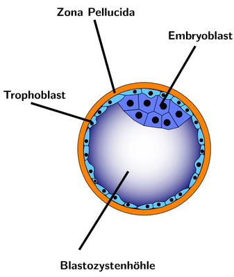
The pluripotent stem cells inside the blastocyst from which the embryo develops are called embryoblasts (see figure). According to the decree of the Law of Regulation of Preimplantation Genetic Diagnosis in December 2011, the examination of cells of the embryoblast is permitted in special cases.
Cells in the blastocyst stage of the embryonic development which form the outer layer of the blastocyst (see figure), are called trophoblast. This layer of pluripotent cells provides the embryo with nutrition and mostly develops to become a placenta. The Trophoblast biopsy in blastocyst stages is a late form of PGD; when reaching this stage, the embryos are 5-6 days old. This is one of the reasons why it was regarded critically for a long time. There is not much time for diagnosis as the embryos are transferred into the mother’s uterus not later than day 6. This temporal pressure can be reduced by shock freezing (cryopreservation) the embryos, because cryopreservation or vitrification is more and more suitable for use.
The acquittal of the Berlin physician through the German Federal Court (BGH) in July 2010, in the course of which the revision of the Embryo Protection Law regarding PGD was induced, refers to the procedure of the trophoblast biopsy.